Microtia is a congenital anomaly of external ear (pinna) that ranges from mild structural abnormalities to complete absence of ear. When an external ear is absence, it is referred to as anotia. Microtia may occurs as an isolated deformity although it can also present itself as part of a spectrum of other deformities, either minor or major such as hemifacial microsomia, Goldenhar syndrome and Treacher-Collins syndrome. Most patients with the most severe form of microtia also lack an external auditory canal, also known as “atresia”, that often require treatment for hearing impairment and surgical ear reconstruction.
The reported prevalence of microtia varies among regions, and it is considered to be higher in Hispanics, Asians, Native Americans, and Andeans. The incidence of microtia is reported to be 1 in 8,000 – 10,000 births. Microtia can affect one side only (unilateral) or both sides (bilateral). Microtia occurs more commonly in males and on the right side. Approximately 10 percent may occur on both sides.
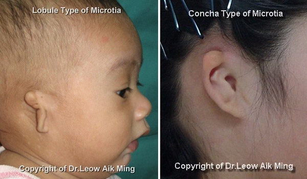
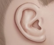
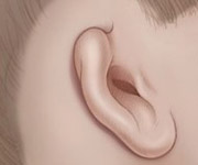
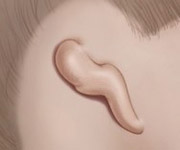
Classification of microtia based on the extent of ear malformation and ear canal damage:

IDEAL CANDIDATES
Factors that may influence eligibility for surgery include:
- Severity of external ear deformity
- Extent of hearing loss
- Age (at least 5-6 years old), preferably 10 years old or the chest circumference at the level of the xiphoid process is to be at least 60 cm.
- General health
- Motivated and having realistic expectations
PRE-OPERATIVE EVALUATION
The decision to undergo reconstructive ear surgery in patient with microtia is personal and should be considered appropriately. Before undergoing reconstructive treatment, surgeon will be working closely with each patient to discuss expectations, preferences, and candidacy for certain procedures.
Not every patient with microtia decides to have reconstructive surgery. Some parents choose to wait and allow their children to decide whether or not to have surgery once they become older. However, younger patients who had successfully undergone ear reconstructive surgery, majority of them regained sense of self-confidence which is essential in the early stage of childhood development.
RECONSTRUCTIVE OPTIONS
The selections of reconstructive option are determined by age, health conditions, patients’ and surgeons’ preferences.
- Prosthetic ear
- Ear reconstruction with synthetic material such as medpor ear implant
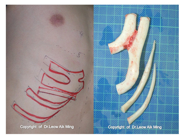
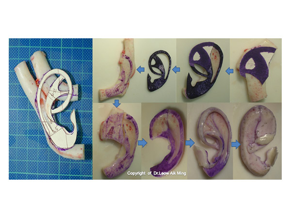
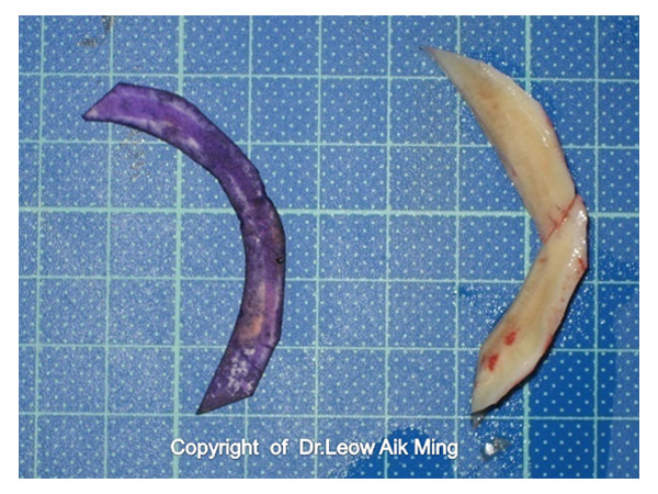
- Autologous reconstruction with rib cartilage (is considered to be the preferred method for microtia reconstruction*)
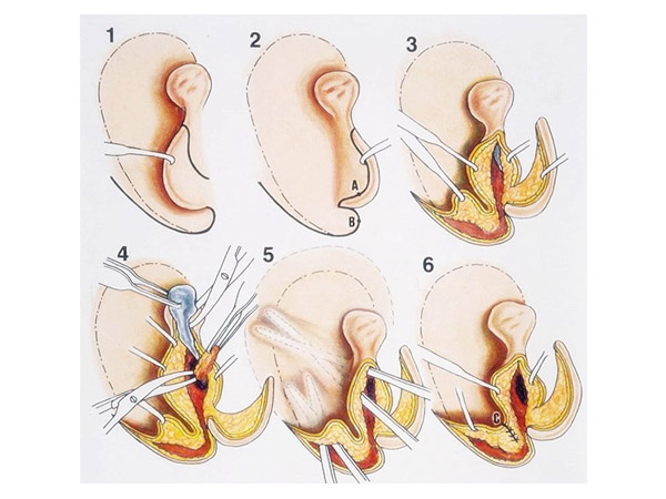
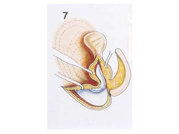
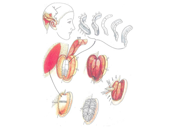
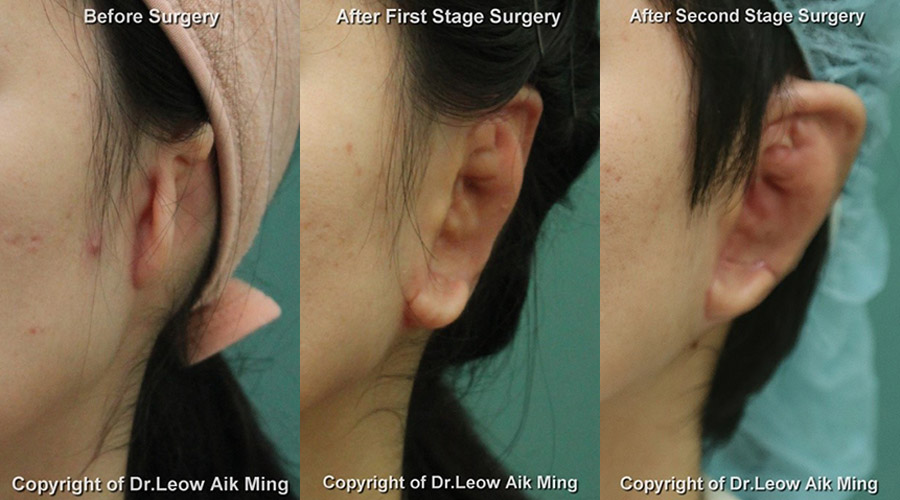
Description of microtia reconstruction with rib cartilage – using two-stage technique:
- A. First stage ear reconstruction
- B. Second stage ear reconstruction
RISKS AND SAFETY
It is important for every parents and patients to understand that every surgical procedure has its own complications and down time. However, if a patient is assessed properly before the surgery and postoperative care is given adequately, these risks can be eliminated or reduced. The risks involved in ear reconstruction for microtia may vary depending upon the nature of the surgical procedure and complexity. Some of the common risks in surgery include:
- Infection
- Hematoma or Seroma
- Skin graft necrosis
- Skin envelope or flap necrosis
- Flap congestion
- Hair loss at operated site
- Cartilage absorption
- Wire or suture extrusion
- Hypertrophy scar
- Unsatisfactory result
- Asymmetry
- Nerve injury
- Complications from donor site such as chest wall deformity, pneumothorax and atelectasis
- Risks of general anaesthesia
POST-OPERATIVE EXPECTATIONS
Reconstructive surgery for microtia of the ear is a complex and challenging surgery. Both parents and patients need to understand the process of recovery and postoperative care to ensure best surgical outcome. Once the reconstructed ear has completely healed, it will not hinder the patient from any activity and will appear completely normal, resistant to any physical stress or strain.
After the surgery, there will be swelling, bruises, numbness, discomfort and pain at the operated sites. Most of the patients do not experience any significant amount of discomfort at any stages of microtia reconstruction. The degree of discomfort or pain varies from one patient to another. Any form of discomfort or pain after surgery can be easily managed with pain medications. The newly reconstructed ear may not be apparent initially after the surgery due to swelling and bruises. It will usually take about a couple of weeks and months for the shape and landmarks of the ear to be clearly visualized. Ear support or protective dressing should be worn at all time during the initial phase of healing to prevent trauma or excessive pressure to the reconstructed ear. Contact sports should be avoided for 4-6 months.
POST-OPERATIVE CARE
- Negative suction drain may be kept for 3-5 days so that the skin can mould to the newly inserted cartilaginous framework
- Soft protective dressing will be applied at all time during early postoperative period until review
- Medications will be prescribed such as antibiotic, anti-swelling pills, pain medication and anti-septic solution
- Sutures are to be removed after 7-10 days
- Wound hygiene should be maintained at all time including washing hair regularly
- Patient can participate light activities with ear protection gear or dressing after 10-14 days
- Contact sports are restricted for about 4 to 6 weeks.
– COPYRIGHT OF DR LEOW AIK MING
